4 New Research Publications with NanoFCM

October 4 2023, Science Advances. DOI: 10.1126/sciadv.adi1556
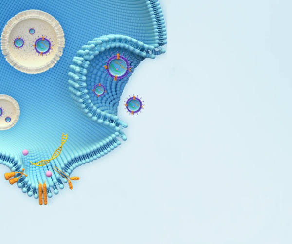
The clinical potential of miRNA-based liquid biopsy has been largely limited by the heterogeneous sources in plasma and tedious assay processes. Here, we develop a precise and robust one-pot assay called dual-surface-protein-guided orthogonal recognition of tumor-derived exosomes and in situ profiling of microRNAs (SORTER) to detect tumor-derived exosomal miRNAs and enhance the diagnostic accuracy of prostate cancer (PCa). The SORTER uses two allosteric aptamers against exosomal marker CD63 and tumor marker EpCAM to create an orthogonal labeling barcode and achieve selective sorting of tumor-specific exosome subtypes. Furthermore, the labeled barcode on tumor-derived exosomes initiated targeted membrane fusion with liposome probes to import miRNA detection reagents, enabling in situ sensitive profiling of tumor-derived exosomal miRNAs. With a signature of six miRNAs, SORTER differentiated PCa and benign prostatic hyperplasia with an accuracy of 100%. Notably, the diagnostic accuracy reached 90.6% in the classification of metastatic and nonmetastatic PCa. We envision that the SORTER will promote the clinical adaptability of miRNA-based liquid biopsy.
August 2 2023, Advanced Healthcare Materials. DOI: 10.1002/adhm.202301706
Extracellular vesicles (EVs) are increasingly being analyzed by flow cytometry. Yet their minuscule size and low refractive index cause the scatter intensity of most EVs to fall below the detection limit of most flow cytometers. A new class of devices, known as spectral flow analyzers, are becoming standards in cell phenotyping studies, largely due to their unique capacity to detect a vast panel of markers with higher sensitivity for light scatter detection. Another class of devices, known as nano-analyzers, provides high-resolution detection of sub-micron-sized particles. Here, the EV phenotyping performance between the Aurora (Cytek) spectral cell analyzer and the NanoFCM (nFCM) nanoflow analyzer are compared. These two devices are specifically chosen given their lead in becoming gold standards in their respective fields. Immune cell-derived EVs remain poorly characterized despite their clinical potential. Therefore, B- and T-cell line-derived EVs and donor-matched human biofluid-derived EVs from plasma, urine, and saliva are used in combination with a panel of established immune markers for this comparative study. A comparative evaluation of both cytometry platforms is performed, discussing their potential and suitability for different applications. It is found that nFCM can accurately i) analyze small EVs (40-200 nm) matching the size accuracy of electron microscopy; ii) measure the concentration of a single EV particle per volume; iii) identify underrepresented EV marker subsets; and iv) provide co-localization of EV surface markers. It can also be shown that human sample biofluids have unique EV marker signatures that can have future clinical relevance. Finally, nFCM and Aurora have their unique strength, preferred fashion of data acquisition, and visualization to fit different research interests.
March 14 2020, Medical Oncology. DOI: 10.1007/s12032-020-1346-1
Renal cell carcinoma is a lethal disease that is often discovered incidentally. New non-invasive biomarkers are needed to aid diagnosis and treatment. Extracellular vesicles (EVs), membranous vesicles secreted by all cells, are a promising potential source for cancer biomarkers, but new methods are required that are both sensitive and specific for cancer identification. We have developed an EV isolation protocol optimized for kidney tumor and normal kidney tissue that yields a high vesicle concentration, confirmed by nanoparticle tracking analysis (NanoSight) and by nanoscale flow cytometry (NanoFCM). Using Western blot, we confirmed presence of EV markers CD81, CD63, flotillin-1, and absence of cellular debris, calnexin. Transmission electron microscopy images demonstrate intact membranous EVs. This new method improves existing protocols with additional steps to reduce contaminants in the EV product. Characterization of our isolation product confirms successful isolation of EVs with minimal contamination. The particle yields of our protocol are consistent and high as assessed by both standard and novel methods. This optimized protocol will contribute to biomarker discovery and biological studies of EVs in renal cancer.
January 10 2022, BMC Cancer, DOI: 10.1186/s12885-021-08870-w
Breast cancer is the most common cancer, and the leading cause of cancer-related deaths, among females world-wide. Recent research suggests that extracellular vesicles (EVs) play a major role in the development of breast cancer metastasis. Axillary lymph node dissection (ALND) is a procedure in patients with known lymph node metastases, and after surgery large amounts of serous fluid are produced from the axilla. The overall aim was to isolate and characterize EVs from axillary serous fluid, and more specifically to determine if potential breast cancer biomarkers could be identified.
